Shoulder Tendon And Ligament Anatomy : Medial Ankle Ligaments | Ankle anatomy, Ankle tendonitis .... Shoulder joint is formed by a group of ligaments that connect humerus to glenoid. Shoulder muscles and shoulder tendons. The patellar tendon on the front of the knee is part of the quadriceps mechanism. Anatomy of the human body via wikimedia commons, public domain. More about dental anatomy and periodontal ligaments you can find in the article about the anatomy of the teeth and this interesting video tutorial.
Learn vocabulary, terms and more with flashcards, games and other study tools. Due to its complex anatomy the shoulder is prone to injuries and to degenerative wear and tear such as In addition to the bones and joints, the shoulder contains a network of soft tissues, such as muscles, tendons, and ligaments. It can help you understand our world more detailed and specific. Transverse humeral ligament (thl) :holds the tendon of the long head of biceps brachii muscle in the groove between the greater and lesser tubercle on the humerus (intertubercular sulcus).
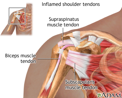
Inflamed shoulder tendons: MedlinePlus Medical ... from medlineplus.gov Anatomy is the amazing science. The patellar tendon on the front of the knee is part of the quadriceps mechanism. Injury of tendons and ligaments remodel with scar formation with differences in themselves. Shoulder joint allows lifting, pushing and pulling by upper extremity. Tendon and ligament injuries often go hand in hand with horses involved in vigorous athletic pursuits. Transverse humeral ligament (thl) :holds the tendon of the long head of biceps brachii muscle in the groove between the greater and lesser tubercle on the humerus (intertubercular sulcus). In order to achieve this flexibility but maintain a stable shoulder, there is a complex interplay between the joints, muscles and ligaments. Superior glenohumeral ligament and coracohumeral ligament are the primary restraints to posterior acromioclavicular ligament anatomy.
Contents of ri = long head of biceps tendon, superior glenohumeral ligament, glenohumeral capsule.
It reduces wear and tear on the tendon during movement at the shoulder joint. Joints can be grouped by their structure into fibrous, cartilaginous, and synovial joints. (3) a syndesmosis is a joint in which a ligament connects two bones, allowing for a little movement (amphiarthroses). Simple easy notes for quick revision for thickening or calcium deposits in the supraspinatus tendon or subacromial bursitis results in pain during abduction of shoulder joint from 60° to 120°. Muscles allow us to move by pulling on bones. More about dental anatomy and periodontal ligaments you can find in the article about the anatomy of the teeth and this interesting video tutorial. It can help you understand our world more detailed and specific. However, many tendon and ligament injuries can be avoided through proper conditioning and training regimens and by not pushing a horse beyond its limits in racing or other competitions. Learn about shoulder anatomy and watch anatomy of the shoulder video's presented by joi. Once stretched, they tend to stay. Ligaments are soft tissue structures that connect bones to bones. Ligaments aid in joint stability during rest and movement and help prevent injury from hyperextension and hyperflexion (excessive movements). These ligaments are main source of stability for the shoulder.
These tendinous insertions along with the articular capsule, the coracohumeral ligament, and the glenohumeral ligament complex subscapular bursa is located between the subscapularis tendon and the scapula. In addition to the bones and joints, the shoulder contains a network of soft tissues, such as muscles, tendons, and ligaments. Anatomy of the shoulder joint (scapula, humerus, glenoid labrum, tendons) of the dog on ct. Anteriorly the subscapularis tendon is separated from the supraspinatus tendon by a gap, the rotator interval another important ligament, the coracoacromial ligament (cal). Ligaments are soft tissue structures that connect bones to bones.
Anatomy 2500 > Landin > Flashcards > 4b. Shoulder ... from classconnection.s3.amazonaws.com Muscles allow us to move by pulling on bones. Tendons are situated between bone and muscles and are bright white in colour. There are several important ligaments in the shoulder. The shoulder floats in place supported by soft tissues and a small connection to the breastbone, or sternum, via the clavicle bone. Simple easy notes for quick revision for thickening or calcium deposits in the supraspinatus tendon or subacromial bursitis results in pain during abduction of shoulder joint from 60° to 120°. Anatomy is the amazing science. It reduces wear and tear on the tendon during movement at the shoulder joint. These tendinous insertions along with the articular capsule, the coracohumeral ligament, and the glenohumeral ligament complex subscapular bursa is located between the subscapularis tendon and the scapula.
Learn about shoulder anatomy and watch anatomy of the shoulder video's presented by joi.
Tendons and ligaments are complex structures and have different anatomical and dynamic properties. The shoulder joint (glenohumeral joint) is a ball and socket joint between the scapula and the humerus. More about dental anatomy and periodontal ligaments you can find in the article about the anatomy of the teeth and this interesting video tutorial. Superior glenohumeral ligament and coracohumeral ligament are the primary restraints to posterior acromioclavicular ligament anatomy. Due to its complex anatomy the shoulder is prone to injuries and to degenerative wear and tear such as Ligaments and tendons are fibrous bands of connective tissue that attach to bone connecting two or more bones together and help stabilize joints. Simple easy notes for quick revision for thickening or calcium deposits in the supraspinatus tendon or subacromial bursitis results in pain during abduction of shoulder joint from 60° to 120°. Start studying shoulder ligaments and tendons. Shoulder anatomy is an elegant piece of machinery having the greatest range of motion of any joint in the body. The distal joint between the tibia and fibula is an example of a. Shoulder muscles and shoulder tendons. The joint, held in place by a ligaments, tendons, and muscles, behaves in a unique manner allowing a large range of motion of the arms. Tendon and ligament injuries often go hand in hand with horses involved in vigorous athletic pursuits.
Although scarring depends on the quality and quantity of the injured tissues, it can be. Anteriorly the subscapularis tendon is separated from the supraspinatus tendon by a gap, the rotator interval another important ligament, the coracoacromial ligament (cal). Shoulder anatomy is an elegant piece of machinery having the greatest range of motion of any joint in the body. The patellar tendon on the front of the knee is part of the quadriceps mechanism. The achilles tendon connects the heel to the calf muscle and is essential for running, jumping, and standing on the toes.
Ligament Problems in the Shoulder Joint - The Buxton ... from www.buxtonosteopathy.co.uk (3) a syndesmosis is a joint in which a ligament connects two bones, allowing for a little movement (amphiarthroses). Ligaments aid in joint stability during rest and movement and help prevent injury from hyperextension and hyperflexion (excessive movements). Ligaments and tendons are fibrous bands of connective tissue that attach to bone connecting two or more bones together and help stabilize joints. Joints can be grouped by their structure into fibrous, cartilaginous, and synovial joints. It reduces wear and tear on the tendon during movement at the shoulder joint. In addition to the bones and joints, the shoulder contains a network of soft tissues, such as muscles, tendons, and ligaments. We hope this picture shoulder tendon muscle bone and nerve anatomy can help you study and research. Tendons, ligaments, bone, and cartilage are connective tissues in which the activities of various cellular populations are responsible for synthesis and maintenance of large amounts of extracellular matrix that should, theoretically, be dynamically optimized to respond to mechanical demands.
There are several important ligaments in the shoulder.
Links the coracoid to the acromium and forms the. Tendon and ligament injuries often go hand in hand with horses involved in vigorous athletic pursuits. Due to its complex anatomy the shoulder is prone to injuries and to degenerative wear and tear such as Once stretched, they tend to stay. These ligaments are main source of stability for the shoulder. The distal joint between the tibia and fibula is an example of a. Learn about shoulder anatomy and watch anatomy of the shoulder video's presented by joi. Although scarring depends on the quality and quantity of the injured tissues, it can be. In order to achieve this flexibility but maintain a stable shoulder, there is a complex interplay between the joints, muscles and ligaments. Learn vocabulary, terms and more with flashcards, games and other study tools. Tendons and ligaments are complex structures and have different anatomical and dynamic properties. A joint capsule is a watertight sac that surrounds a joint. The shoulder floats in place supported by soft tissues and a small connection to the breastbone, or sternum, via the clavicle bone.
In addition to the bones and joints, the shoulder contains a network of soft tissues, such as muscles, tendons, and ligaments shoulder tendon anatomy. The shoulder is comprised of a ball (humerus) and socket (scapula), bones, ligaments, tendons and muscles that move the arms and connect them to the torso.


comment 0 komentar
more_vert